Why Your Knee Pain Isn’t Just About the Knee: Understanding Plica Syndrome Through a Whole-Body Lens
Most people have never heard of a “plica,” let alone know that it could be the hidden reason behind their chronic joint pain. Even among clinicians, plica syndrome is often misunderstood, misdiagnosed, or dismissed as incidental. And when it is treated, the focus is almost always too narrow—centered on the joint, not the body behind it.
At Movability, we’ve built a reputation on solving cases like these—when symptoms seem to have no clear cause, and conventional care has failed. One of the most overlooked contributors we see in chronic joint pain, especially in the knee, is an inflamed or overloaded plica—a small, embryological remnant of synovial tissue that can have big consequences when irritated.
But here’s the most important point:
The plica isn’t the problem—it’s the victim.
Until you find and fix what’s really overloading it, the pain will come back. That’s where we come in.
What Is a Plica? The Science, the Anatomy, and the Forgotten Fold
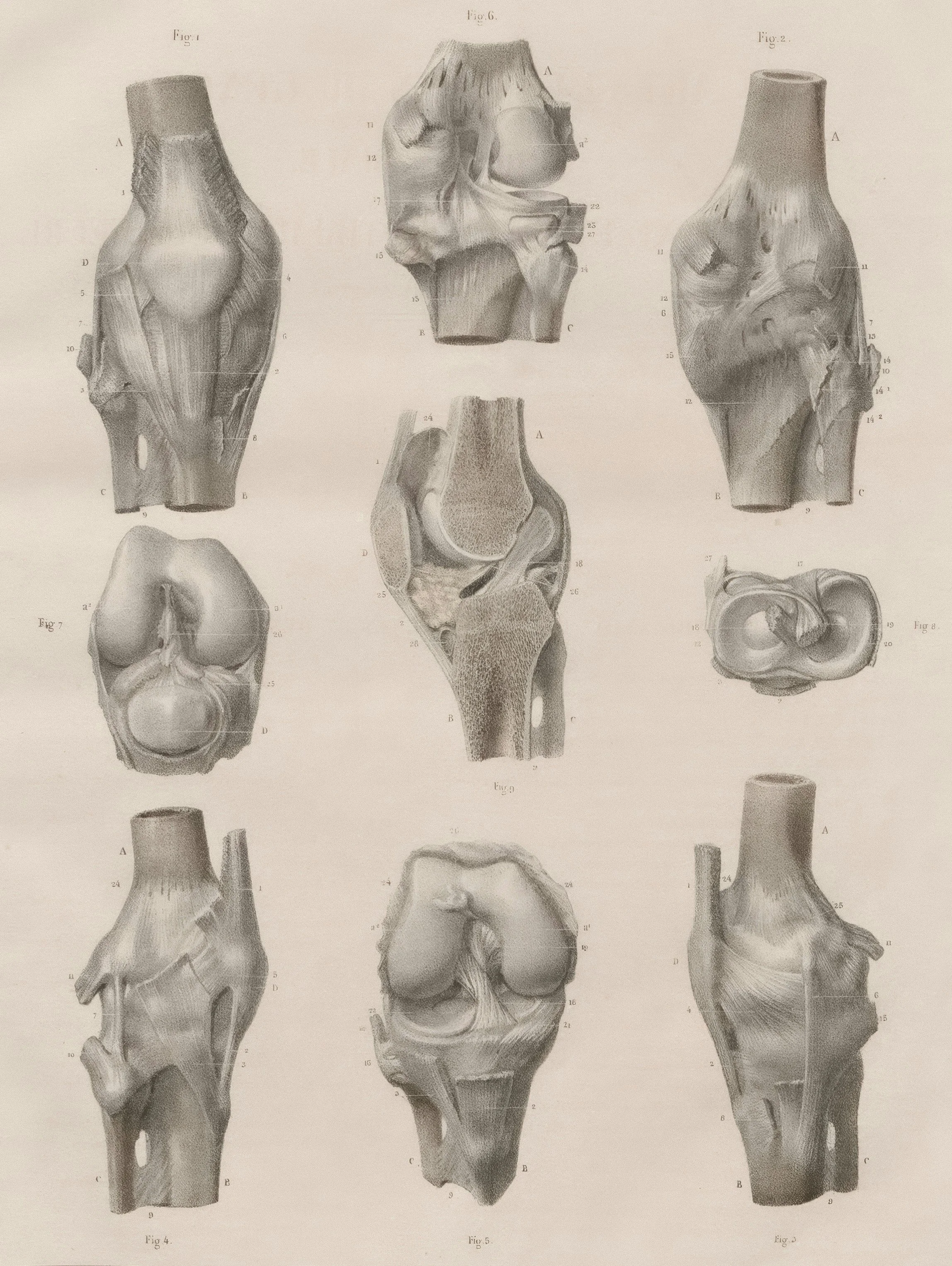
The term plica (Latin for “fold”) refers to synovial membrane remnants left over from fetal development. In the womb, our joints begin as multiple separate cavities, divided by septa. Over time, these cavities merge—but when they don’t fully fuse, thin, elastic folds of synovial tissue can remain. These are called synovial plicae.
These folds exist in many joints:
Knee (most common): suprapatellar, infrapatellar, lateral, and mediopatellar plicae
Elbow: radiocapitellar plica
Ankle: anterolateral meniscoid lesions
Wrist: dorsal capsular or midcarpal synovial folds
Shoulder: subacromial or glenohumeral plica
Temporomandibular joint (TMJ): emerging evidence of synovial folds in some cases
While these plicae are usually asymptomatic, mechanical overload, trauma, or chronic compensation can inflame and thicken them, leading to what’s known as plica syndrome.
Knee Plica Syndrome: The Classic Case, Misunderstood
The mediopatellar plica is the most common culprit. It runs from the medial capsule, across the medial femoral condyle, toward the fat pad. In a healthy knee, it’s pliable and inconsequential. But when it’s chronically compressed—especially between the patella and femur—it loses elasticity and becomes fibrotic, thickened, and pain-sensitive.
Patients often present with:
Intermittent medial knee pain
Snapping, catching, or clicking with knee flexion (usually around 30–70 degrees)
Pain after sitting (positive “movie theatre sign”)
Sensitivity on the inside of the patella
No clear findings on MRI or X-ray
It’s often misdiagnosed as:
Patellofemoral pain syndrome
Meniscal tear
Chondromalacia
Early arthritis
The Real Reason the Plica Gets Irritated: Pelvic Obliquity, Gait Changes, and Neuromuscular Imbalance
The plica is rarely the root cause.
At Movability, we’ve learned that it’s a warning sign of deeper biomechanical dysfunction.
The Case of the Marathon Runner Who Did Everything Right
She was 42, disciplined, and fast. A seasoned marathon runner who had logged thousands of miles over the years without so much as a twinge in her knees. She ate clean. Trained smart. Had a coach. Rotated her shoes. Switched terrain. Stretched. Recovered.
So when her right knee started hurting, she couldn’t believe it.
At first, it was just a faint ache on the inner part of the knee after a long run. Nothing major. Then it started catching slightly on stairs. The discomfort became sharp at times, especially after sitting. She saw a practitioner who diagnosed her with patellofemoral pain syndrome and gave her exercises to “strengthen her quads.”
But the pain kept coming back. Worse some days. Completely absent on others. She was doing everything right — and yet, she was getting worse.
That’s when she came to Movability.
Dr. Sina met her for an assessment. “You’re clearly experienced,” he said. “You know your body. If you say something feels off, I believe you.”
He ran her through a full head-to-toe exam — manual muscle testing, posture screening, gait analysis, balance control. That’s when he picked up on a telltale sign: a thickened medial plica on the right knee. It was snapping over the joint with flexion and inflamed on palpation. She had classic plica syndrome — something that had gone completely missed.
“But why now?” she asked. “Why would this happen after all these years of injury-free running?”
That’s when Dr. Sina asked one of the most important questions in the entire appointment:
“Has anything changed recently in your life — not just in training, but your environment? Your work setup? Sleeping position? Anything you wouldn’t even think is connected?”
She paused. Then her eyes lit up.
“Well… I did renovate my office,” she said. “I got a new corner desk a few months ago. My monitor sits off to the side now. I guess I sit a little twisted all day.”
That was the moment the real diagnosis clicked into place.
After months of sitting slightly rotated, her body had adapted — but not in a good way. She’d developed pelvic obliquity, with her right pelvis slightly lower than her left. This made her right leg functionally longer. The result? Every step she ran loaded the right knee just a bit more. The angle of her femur changed. Her gait subtly compensated. Over thousands of strides, the medial plica was slowly but consistently being overloaded — until it finally snapped.
But there was one more layer.
Her eyes, constantly focused off-center, had trained her nervous system into a new postural default. Her cervical spine, visual reflexes, and vestibular system had adapted to this asymmetry — reinforcing the imbalance all the way down the chain.
It wasn’t just a knee problem. It was a full-body compensation that started with her desk chair.
Together, Dr. Sina and the Movability team built a treatment plan that included:
Manual therapy to release the plica and surrounding tension
Pelvic realignment to correct the obliquity
Neuromuscular retraining for her gait and hip firing
Vestibular and gaze stabilization to recalibrate her postural reflexes
Ergonomic coaching to fix her workstation once and for all
She didn’t need surgery. She didn’t need another misdiagnosis.
She needed someone to ask the right question.
Had we simply treated the knee, she would have been back in 6 months with the same problem—or worse.
Neurological and Sensory Drivers of Plica Overload
Recent studies show that pathologic plicae have:
High concentrations of Substance P-positive pain fibers
Increased vascularization and mechanoreceptors
Evidence of neurogenic inflammation
This means the plica is not just an innocent bystander—it’s a highly innervated structure that becomes reactive under stress. When joint alignment, gaze orientation, or vestibular input becomes chronically altered, this stress is funneled toward mechanical hotspots—often the plica.
That’s why eye position, vestibular reflexes, and cervical posture all influence how the knees behave. At Movability, we account for this full sensory-motor network, not just joint angles.
Other Plicae in the Body: Why You Shouldn’t Only Think of the Knee
Though knee plica syndrome is the most common, other joints can suffer from the same problem:
Elbow (Radiocapitellar Plica Syndrome)
Lateral elbow pain mistaken for tennis elbow
Clicking or snapping at terminal extension
Seen in throwers and manual workers
Wrist Plica
Pain with wrist extension (e.g. in yoga or push-ups)
Often found in the dorsal capsule or scaphotrapeziotrapezoid joint
Easily missed on imaging
Ankle Plica
Post-sprain anterolateral pain
Mimics impingement or instability
May present with catching or limited dorsiflexion
Shoulder and TMJ Plicae
Rare, but when present, can mimic impingement or labral pathology
May contribute to persistent pain in high-functioning athletes or postural dysfunctions
Why Conventional Treatment Fails: The Plica Is the Messenger, Not the Villain
Most conventional approaches to plica syndrome include:
Rest, NSAIDs, taping
Corticosteroid injections
Arthroscopic plica resection
These may reduce inflammation temporarily, but don’t address why the plica became overloaded. That’s why recurrence rates can be high, especially in active individuals.
At Movability, we’ve found that when we restore full-body balance, the plica doesn’t just settle—it stays calm.
The Movability Method: Root-Cause, Collaborative, and Results-Driven
Our clinic specializes in complex, recurring, and misdiagnosed cases. We don’t chase symptoms—we reverse engineer dysfunction from the ground up.
Our Process Includes:
Detailed movement screening
Pelvic and postural alignment testing
Cervico-ocular and vestibular input assessment
Manual orthopedic testing to pinpoint involved structures
3D gait and foot pressure analysis
Collaborative review with physiotherapy, chiropractic, and naturopathic teams
Treatment Is Personalized and Layered:
Manual therapy for the plica and compensating structures
Pelvic and spinal realignment
Neuromuscular re-education for functional gait and posture
Vestibular retraining to recalibrate the entire kinetic chain
Corrective exercises and load management
Nutritional and anti-inflammatory support through our naturopath
We don’t do cookie-cutter rehab—we build a precision-matched plan for your body and lifestyle.
Long-Term Outlook and Prevention
When treated correctly, the long-term outlook for plica syndrome is excellent. Once the underlying asymmetry, postural driver, or neuromuscular imbalance is corrected, recurrence is rare. Many of our patients report not only resolution of their knee pain but also improvements in balance, performance, and body awareness.
We equip each patient with the tools to maintain results through:
Ergonomic education
Custom home exercises
Follow-up strategies to reinforce new movement patterns
Unlike treatments that only target the symptom, our integrative care prevents the problem from coming back.
Frequently Asked Questions
What are the symptoms of plica syndrome?
Pain on the inside of the knee, snapping or clicking with motion, pain after sitting, and sensitivity near the patella. Some patients also experience catching or a “giving way” sensation.
Can you see a plica on MRI?
Sometimes, but not always. Many plicae are too thin to be visualized unless they’re inflamed or fibrotic. A skilled physical exam often reveals more than imaging.
Is surgery required for plica syndrome?
Not always. In fact, most cases resolve without surgery if the root cause—like postural imbalance or pelvic obliquity—is addressed properly.
Can plica syndrome occur in other joints besides the knee?
Yes. Plica-like folds have been found and documented in the elbow, ankle, wrist, shoulder, and even the TMJ. When inflamed, they can cause joint-specific symptoms.
Why does plica syndrome come and go?
Because the mechanical irritation may depend on posture, activity, or stress levels. Unless the underlying dysfunction is fixed, the plica will continue to flare intermittently.
Our Promise: Treat the Source, Not Just the Site
Whether it’s your knee, elbow, ankle, or wrist—if you’ve been told there’s “nothing wrong,” or you’ve had short-term success followed by repeat flare-ups, plica syndrome might be the missing link.
But more importantly, the plica might be trying to tell you something about your posture, your balance, your ergonomics, or your nervous system.
At Movability, we listen.
Contact Movability today to book a comprehensive assessment and experience our results-driven approach.